Is it time for your annual comprehensive eye exam? Most adults have experienced an annual eye exam, and understand that the exam accomplishes more than just checking for 20/20 vision: Regular eye exams help protect your overall eye health. Whether it’s your or your child’s first exam, knowing what to expect makes the experience easier.
Eye exams — which are comfortable and non-invasive — provide your eye doctor with two important types of information:
- How well you are seeing and whether you need vision correction (or a change in your prescription)
- The overall health of your eyes and visual system
If your child is coming in for their first comprehensive eye exam, reassure them that the exam won’t hurt, and that the eye doctor will answer their questions during the exam.
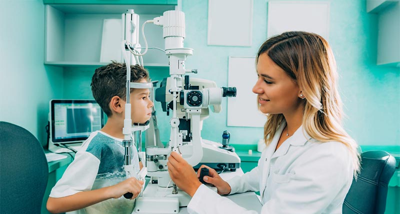
Regular eye exams should begin between ages 3 and 5 to establish a visual baseline. With today’s diagnostic equipment, your child doesn’t even need to know how to read or recognize the letters of the alphabet to have their eyes examined!
Step-by-Step: What Happens During the Exam
Our trained staff and doctors will guide you through a comprehensive series of tests to provide an overall assessment of your visual health. These tests are quick and painless, and provide a wealth of information about your visual system. Following is a general overview:
- Patient Visual and Medical History. We’ll ask about any vision changes or any changes in your general health, medications, and work or environmental conditions that may affect your vision. Your family medical history is important. Discuss with your doctor any personal and family history of high blood pressure, diabetes, autoimmune disease, thyroid, or other systemic disease.
- Visual Acuity. This test uses the Snellen Eye Chart — that cool-looking chart with the big E at the top and rows of letters that gradually get smaller below it. We use it to measure your visual acuity, meaning how clearly you see both at a distance and up close. Your visual acuity is based on the smallest row of Snellen Eye Chart letters that you can read clearly from 20 feet away. We’ll also test your up-close vision with a small card held about 14 inches away from your eyes.
- Visual Refraction. An instrument called a phoropter helps determine the exact prescription lens power you need to achieve 20/20 vision. The phoropter incorporates various lenses and lens power combinations. Your eye doctor will change these combinations to pinpoint the exact prescription correction to compensate for your nearsightedness, farsightedness, astigmatism, or any combination of the three. During refraction, your optometrist may ask, “Which is better: 1 or 2? 3 or 4?” The test incorporates your input to determine which lenses give you the clearest, most comfortable vision.
- Eye Muscle and Alignment Tests. Your optometrist will check for eye teaming, focusing, and binocular vision abilities. This measures how effectively your eyes can focus, move, and work together in unison to help you perceive the world around you.
- Visual Field Test (Perimetry). We will measure your central and peripheral vision to detect any blind spots that could be linked to glaucoma, stroke, diabetes, or optic nerve-related conditions.
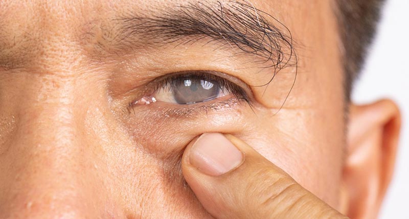
- Slit Lamp Examination. The slit lamp is a microscope with a bright light. It allows your optometrist to examine your eyelids, eyelashes, and other external tissues, as well as the sclera (the whites of your eye), conjunctiva, cornea, iris, pupil, and lens. A slit lamp exam helps the doctor assess your overall visual system health and assists in detecting cataracts, dry eye, and corneal disease, among other conditions.
- Eye Pressure Test (Tonometry). When you rest your chin on this device, it delivers a small puff of air that measures intraocular pressure to screen for glaucoma.
- Imaging Tests. A picture is worth a thousand words and can help save your vision. Depending on your age and health issues, your doctor may take photos of your visual system. These may include:
- Retinal imaging: A detailed picture of the back of the eye using a special camera. This photo captures details of your retina, optic nerve, and blood vessels to assist in detecting visual changes related to diabetes, hypertension, and macular degeneration.
- Optical Coherence Tomography (OCT): This imaging device uses light waves to create a cross-section of the retina that can allow your doctor to see early signs of diseases, including glaucoma, macular degeneration, or retinal swelling.
- Dilation. When needed, your eye doctor may use eye drops that enlarge your pupils to gain a better view into the back of the eye to examine its critical structures. Dilation can help diagnose retinal tears, retinal detachments, tumors, and other signs of systemic disease. Dilation onset takes between 15 and 20 minutes and can last for several hours. Dilation can cause light sensitivity, trouble focusing, or blurry vision in some patients, so plan accordingly.
While your eye doctor may add other tests depending on your age and stage of life, these nine tests are typical during a comprehensive eye exam to measure and evaluate overall eye health and visual acuity. Each individual test provides details about a specific facet of your eye health. Depending on your eye health, age, and medical history, your optometrist may or may not administer all of these tests every year.
Early detection and treatment of vision challenges help prevent vision loss. That’s why it’s so important to have an annual comprehensive eye exam, and to schedule one for your children.
Call our office today to schedule your exam and let us check out your “windows on the world” to ensure you’re seeing your best.